Give a blood cell a 3.75 m width; DNA becomes 1 mm wide
A red blood cell’s width and a DNA double helix’s width are very different. First express both in nanometres. Then the comparison is ordinary division.
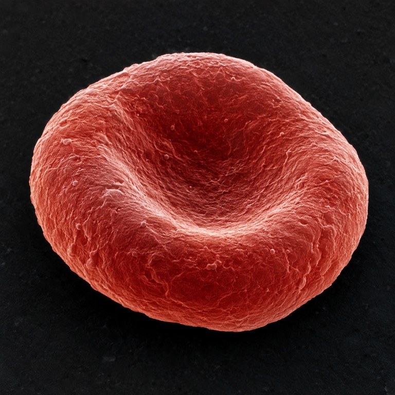
Work it out, one step at a time
Use the same unit
7.5 µm × 1,000 = 7,500 nm
One micrometre contains 1,000 nanometres.
Divide the larger width by the smaller
7,500 ÷ 2 = 3,750
This is a width ratio. It does not count how many DNA molecules fit inside a cell.
Words made simple
- Diameter
- The width across an object through its centre.
Think about it: If DNA is 2 mm wide in your model, how wide is the cell?
2 × 3,750 = 7,500 mm, or 7.5 m. Both lengths must grow together.
Where the numbers come from: NHGRI · Fung, Tsang & Patitucci (1981). The illustration and worked model are made for this lesson; assumptions are stated above.
Go further: definitions, formulas and a second example
A cell can be microscopic and still be thousands of times wider than a molecular structure. Taking the ratio is easy; understanding what the numerator and denominator mean is the scientific part of the comparison.
A cell diameter and a molecular width
ScaleTrip uses 7.5 µm as a representative red-blood-cell diameter and about 2 nm for DNA’s double-helix width. The cell value is a chosen comparison, not a universal constant. The linked geometry study reported a different sample mean and describes biological variation.
The DNA number describes width across the helix, not the full contour length of a DNA molecule. A long thin thread can be narrow and long at once. Keeping “diameter” or “width” next to both records avoids accidentally comparing cell width with the total length of genetic material.
Normalize the units before dividing
One micrometre is one thousand nanometres, so 7.5 µm becomes 7,500 nm. Dividing 7,500 by 2 gives 3,750. This is a dimensionless length ratio: the chosen cell diameter is 3,750 times the chosen DNA width. The representative inputs set the meaning and precision of the result.
A one-metre model cell would give that model DNA a width of about 0.267 mm. The arithmetic is 1 m / 3,750. It is a scaled-width comparison, not a model showing how DNA is arranged inside a red blood cell or any claim that these objects have the same internal architecture.
A length ratio is not a packing count
Cubing 3,750 would give the volume ratio of geometrically similar objects scaled in all three dimensions. A biconcave cell and a double helix are not such a pair. The length result alone therefore cannot tell you how many DNA molecules fit in a cell or what fraction of its volume they occupy.
The same care applies to the image. Enlarged DNA rungs and a cell outline may be displayed at similar screen widths to remain recognisable. The numerical ratio remains useful precisely because it is calculated from the stated lengths rather than inferred from the displayed artwork.
Turn the comparison into a measurement question
Select another cell diameter in the comparison tool and predict whether the ratio increases proportionally. If the diameter changes by ten percent while DNA width stays fixed, the linear ratio changes by ten percent too. This is a transparent sensitivity test, not a biological experiment.
End by naming both measurements, reporting the representative nature of the cell value, and limiting the conclusion to width. That short explanation is more informative than a dramatic zoom alone, because another reader can repeat it and see exactly what would change with new data.
Reproduce one result
Calculate this result using the definitions and assumptions in the article.
- Expression
- (7.5 × 10⁻⁶) / (2 × 10⁻⁹)
- Calculated result
- 3,750 ×
Check the numbers and their sources
Open a source to see what it supports. The picture is our explanation; the original evidence is linked below. These AI-assisted explanations have automated calculation checks, but no expert peer review.
Fung, Tsang & Patitucci (1981) — Red blood cell geometry (abstract)
Publisher abstract only; full article restricted. ScaleTrip keeps a chosen 7.5 µm representative and illustrative 6–8 µm band, not universal bounds. Source record checked:
Read the original sourceNHGRI — DNA Day transcript, question 296
Diameter, not the length of a DNA molecule. Source record checked:
Read the original sourceNIST — Metric (SI) prefixes
A unit definition is not a measured object size. Source record checked:
Read the original source